 Aspiration is when a dog or cat inhales food, liquid, or stomach contents down their airway (trachea) instead of swallowing down their esophagus.
Most of us have experienced this at some point in our lives when a small amount of liquid goes into our windpipe and causes us to cough and feel uncomfortable for a short period.
Some pets may cough when an aspiration event occurs, but some pets do not, called “silent aspiration,” which can make it difficult to know that there might be a problem.
Aspiration pneumonia develops when the material swallowed into the lungs causes inflammation or infection.
This condition seems to affect dogs more than cats, but it can happen to any pet. See below for the conditions that can cause aspiration.
Is Aspiration Pneumonia Serious?
The lungs contain very small air sacs called alveoli. The alveoli are the only place in the body where oxygen exchange for carbon dioxide occurs – a vital life-sustaining process. When a large amount of foreign material clogs these tiny air sacs, it causes difficulty breathing and significant inflammation or infection, leading to pneumonia.
If you suspect your pet has swallowed something into their airway, pay close attention, monitor them over the next few days, and get them to a vet as soon as possible if the symptoms listed below emerge. An infection can develop quickly, especially from swallowing food or stomach contents (vomit) into the trachea.
What Are the Symptoms of Aspirating?
- Coughing
- Lethargy
- Not eating well
- Coughing up phlegm*
- Fever (temperature greater than 103.5°F)*
- Labored Breathing – breathing with extra effort and/or greater than 40 breaths per minute*
- Difficulty getting up*
- Unable to get up*
- Pale, blue, or purple gums*
*If your pet has these signs, please go to a veterinarian immediately!
Daisy, a toy poodle of one of our Preventive Vet team members, aspirated in the middle of the night while she was sleeping. Right away, her family noticed that she wasn't breathing well and rushed her to the emergency hospital within 30 minutes.
At the ER, the physical exam and X-rays (seen further in this article) determined she had indeed aspirated and that an infection (pneumonia) had started. She spent two days in the hospital recovering.
In the video below, Daisy is breathing with extra effort and a noisy wheeze when she exhales. Her respiratory rate is 44 breaths per minute. Breathing with this much effort is exhausting, as the average breathing rate for healthy dogs and cats is between 15 and 30 beats per minute. Wheezing and heavy breathing indicate that Daisy needs immediate veterinary care.
Can Any Dog or Cat Aspirate?
Typically, aspiration does not happen in normal, otherwise healthy animals. Dogs and cats who are awake and feeling well do not have trouble swallowing food or water. If some food or water happens to go down the wrong way, it is usually a small amount that they can cough out.
However, there are certain medical issues that increase the chance for a cat or dog to aspirate.
Medical Conditions and Circumstances That Could Cause Aspiration
Sedation
Pets who are sedated or anesthetized are extremely relaxed. This relaxation often causes a loss of the protective swallow reflex. It may also cause the sphincter that keeps stomach fluids inside the stomach to relax, allowing stomach fluid to flow toward the mouth. This creates a risk for aspiration.
This is one big reason that you don't feed your pet before a dental cleaning, and they are often given an anti-nausea medication (often Cerenia®) so they won't vomit upon coming out of anesthesia.
Seizures
A pet with a severe seizure may also temporarily lose that protective swallow reflex. Aspiration pneumonia is a rare complication of a seizure.
Bottle-fed pets
Pets who are syringe-fed or bottle-fed food or medications may inhale the material into their lungs. This is especially common with sick newborn pets with weaker swallow reflexes.
Diseases and disorders that affect swallowing
Pets who have difficulty swallowing are at risk of aspiration pneumonia because they may inhale food or water. Diseases that may cause swallowing difficulties include:- Laryngeal paralysis – Animals have gates called arytenoids in their throat, which close when they eat. These gates prevent food and water from passing into the airway. In laryngeal paralysis, one or both gates are partially paralyzed, so food or water can be inhaled anytime the pet eats or drinks. Severe laryngeal paralysis may be addressed with surgery.
- Cricopharyngeal achalasia, cricopharyngeal asynchrony – In cricopharyngeal achalasia, the muscles of the throat do not relax enough to allow normal swallowing. In cricopharyngeal asynchrony, the opening and relaxation timing of the throat muscles is not coordinated. Cricopharyngeal achalasia can be treated with surgery to release the tight throat muscles.
- Mass or abscess (pocket of pus) in the throat area – If the throat has a partial blockage, it is possible for a dog or cat to inhale food or water instead of swallowing it. An abscess is treated with surgery. Depending on the type of mass, it may be treated with surgery or with medical therapy.
- Disorders of the esophagus can cause regurgitation, leading to aspiration.
Examples include:
- Megaesophagus – In this disorder, the muscles of the esophagus lose their ability to move food and liquid normally. The esophagus becomes enlarged, and the pet regurgitates frequently. There are numerous underlying causes of this disorder. There are medical treatment options, but dietary management is also quite important.
- Esophagitis – This is an inflammation of the esophagus. It is often due to gastroesophageal reflux (heartburn). But it can also be due to an irritation from a foreign object in the esophagus. Esophagitis may be treated with medication, but a definitive diagnosis requires an endoscope inserted into the throat to view the esophagus from the inside. An endoscopic procedure can also remove a lodged foreign object.
- Esophageal Stricture – A stricture is a narrowing of the esophagus that may occur after a bout of severe esophagitis or due to a lodged foreign object (such as a bone, rawhide, or toy). An endoscopic procedure using special balloons can dilate the esophagus to help widen it again, although it often requires multiple procedures until the pet can eat normally again.
- Megaesophagus – In this disorder, the muscles of the esophagus lose their ability to move food and liquid normally. The esophagus becomes enlarged, and the pet regurgitates frequently. There are numerous underlying causes of this disorder. There are medical treatment options, but dietary management is also quite important.
- Illnesses that cause frequent vomiting.
Examples include:
- Pyloric stenosis – This is a disorder of puppies and kittens who vomit frequently once they start on solid food because they were born with a narrowing at the exit of their stomach. This is diagnosed with a barium study or endoscopy and treated with surgery.
- Stomach or intestinal blockage – Common causes of a gastrointestinal blockage include a pet eating a foreign object that is too large to pass on its own or a mass. Masses may be cancerous or non-cancerous tumors, abscesses (pockets of pus), or granulomas (localized areas of inflammation). This may be diagnosed with X-rays or ultrasound and may be treated with endoscopy or surgery.
-
Inflammatory bowel disease (IBD) – IBD is an abnormal infiltration of immune cells in the stomach or intestine. This can occur in cats or dogs and most commonly occurs in young to middle-aged pets, although older pets can be affected.
Some pets who have severe inflammation in the stomach or upper intestine may vomit frequently, which puts them at risk for aspiration pneumonia. Diagnosis is through a biopsy. Some pets respond to a hypoallergenic diet, while others respond to antibiotics (more often dogs than cats), and the rest do best on immune suppressants such as steroids. -
Parvovirus/Panleukopenia – These are similar viruses that affect dogs (Parvovirus) and cats (Panleukopenia). There are vaccines available for both viruses. However, outbreaks of both viruses are seen in areas where pets and stray animals are not routinely vaccinated. Both viruses lead to vomiting, diarrhea, and a marked decrease in the white blood cell count.
The low white blood cell count can make the pet susceptible to secondary bacterial infections on top of the initial viral infection, making this a life-threatening combination. The vomiting and diarrhea can be quite severe, and aspiration pneumonia is possible.
A pet with vomiting and diarrhea who has not been vaccinated needs immediate veterinary attention to control vomiting, keep them hydrated, and start antibiotic therapy.
- Pyloric stenosis – This is a disorder of puppies and kittens who vomit frequently once they start on solid food because they were born with a narrowing at the exit of their stomach. This is diagnosed with a barium study or endoscopy and treated with surgery.

How a Pet Is Diagnosed and Monitored During Treatment
Physical Examination
Possibly including a neurologic examination since some causes of swallowing difficulties and regurgitation are neurologic.
A neurologic examination assesses the pet’s mental alertness, awareness of the position (called proprioception), and movement of their body (called postural reactions) and checks reflexes to see if the nerve function appears normal.
Finally, a veterinarian feels along the head and spine for any signs of pain or swelling. Together, any abnormal findings help the veterinarian to narrow down the list of potential causes of a neurologic condition.
Chest X-Rays
X-rays are very helpful to see changes in the airways consistent with aspiration pneumonia. In some pets, it can take up to 24 hours after an animal aspirates for pneumonia to be seen on X-rays. Once it is seen, X-rays can be used to monitor treatment progress and healing.
Even if aspiration pneumonia is not seen on initial X-rays, treatment can and should be started as soon as possible for a pet who is having difficulty breathing. Often, a pet who is having difficulty breathing may be started on treatment based on other diagnostics and a history consistent with possible aspiration.
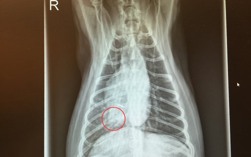
The red circle above outlines the alveolar lung pattern in the right middle lung lobe. The right middle lung lobe is a common lobe where aspiration pneumonia occurs. An alveolar pattern indicates that the alveolar air sacs of the lung are completely clogged with debris.
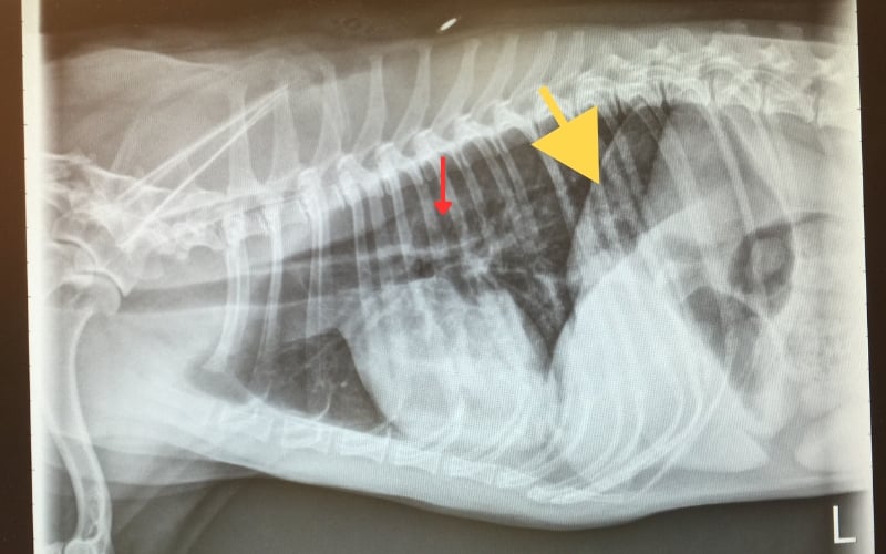
In the X-ray below, the affected lung is lying over the oval-shaped heart, so the pneumonia is difficult to see. The area with the pneumonia appears as bright-white patchy areas over the heart. The small red arrow shows a small dilation of the esophagus.
The wide yellow arrow shows gas in the stomach because Daisy was working so hard to breathe that she was also swallowing extra air. This is common in pets who are struggling to breathe.
Lung Ultrasound
More veterinarians are becoming trained in this technique to diagnose signs of pneumonia using ultrasound. This technique can also be used to monitor healing.
C-Reactive Protein
C-reactive protein is a blood test that measures inflammation in the body. If a pet has aspiration pneumonia and the C-reactive protein levels are decreasing, this is a good sign that the treatment is working.
Complete Blood Cell Count
Pets with aspiration pneumonia may develop increased white blood counts, particularly the neutrophil count. Neutrophils are the white blood cells tasked with fighting bacterial infections.
Pulse Oximetry
This test measures the oxygen saturation of the blood. This non-invasive test detects if a pet is not getting enough oxygen when they breathe.
Arterial Blood Gas
This blood test is taken from an artery instead of a vein. Blood is usually collected from veins, which is blood mostly depleted of oxygen returning to the lungs to exchange its carbon dioxide. Arteries carry oxygenated blood, so this test is a sensitive lung function test.
Collection of Respiratory Samples
Bronchoscopy with bronchoalveolar lavage – This procedure is performed under anesthesia to both visualize the inside of the airway and collect a deep wash sample of cells for culture (to determine the best antibiotic choices) and cytology (for examination under the microscope).
In a bronchoalveolar lavage, a small amount of sterile saline is placed deep into the airway and then immediately retrieved. The sample that is retrieved contains respiratory epithelial and inflammatory cells, as well as infectious organisms, for a pathologist to view under the microscope. The fluid can be (cultured) tested in a lab to see if it grows bacteria. If it does, it will be tested against known antibiotics to see which will ones will work best against this infection.
Transtracheal wash – This test is performed under heavy sedation using a long catheter placed in the trachea to collect a wash sample. The fluid can be used for a pathologist review and culture. This is a very similar procedure to the bronchoalveolar lavage described above, but the sample retrieved may not extend as deep as the pneumonia, so it is not always diagnostic. It is typically reserved for patients who cannot be anesthetized for safety or cost reasons.
Additional tests may be needed to determine why your pet has aspirated food or fluid in the first place.
Treatments for Aspiration Pneumonia
Your veterinarian will determine the best course of treatment based on diagnostic tests and how well your pet is breathing.
Pets who are dehydrated, have difficulty breathing – such as using their abdominal muscles to breathe, extending their neck forward to breathe more easily – or have a low oxygen saturation would benefit from hospitalization.
Hospitalization for Serious Cases May Include:
- Intravenous fluids
- Intravenous antibiotics
- Oxygen supplementation
- Oxygen chamber
- Nasal line
- Oxygen hood over the head
- Face mask
- Flow-by (an air hose in front of the face)
- Intubation with ventilation
- Nebulization
- Repeat X-rays, lung ultrasounds, and lab tests as needed to monitor therapy
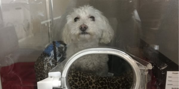
Daisy, in the photo below, who suffered from aspiration pneumonia, spent two days in the ICU, and much of that was in an oxygen chamber to ease her breathing. Her family went to visit her multiple times a day.
With Daisy’s pneumonia, she breathed with more effort and faster to get the oxygen she needed, which made her tire out quickly. By increasing the oxygen concentration in the oxygen chamber, Daisy could breathe more easily because there was extra oxygen in the air. This allowed Daisy to rest while the antibiotics started to work on the lung infection.
More stable cases may be managed at home with:
- Oral antibiotics
- Saline nebulization can be performed at home if your pet tolerates it and your vet recommends it. The procedure uses a nebulizer device like this.
- Keep your pet moving! Movement helps to break up the debris in their lungs and makes it easier for your pet to cough it up. The goal is to remove that infected material rather than let it stay in the lungs. Since the pet may not feel well, short, frequent walks or play sessions will work best initially.
How Aspiration Pneumonia Is Prevented
Before Sedation or Anesthesia
Ensure your pet has not eaten anything. If your pet needs an emergency procedure, let your veterinarian know when your pet last ate so that the staff can take special precautions against aspiration.
 Control Seizures
Pets who have a history of severe or frequent seizures can take medications to prevent or reduce the number of seizures they may have. Your veterinarian can discuss the best medications for your pet based on their history.
Daisy, featured throughout this article, suffered from epilepsy and was on medication that managed her seizures.
She never aspirated due to seizures, as they were well controlled, but she aspirated because she suffered from bouts of regurgitation and she was sleeping when the incident happened.
Vomiting
Prescription anti-nausea medications may slow down vomiting for some pets. However, if your pet is frequently vomiting or the vomiting occurs multiple times a week, a veterinary evaluation for pneumonia is necessary. It's necessary to determine the underlying cause of the vomiting to prevent aspiration events in the future.
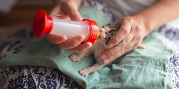
The Proper Way to Bottle-Feed
If you have to bottle-feed newborn puppies or kittens, they should be fed sitting with the back upright (not down like human babies). Feeding them in a human position can lead to aspiration pneumonia. This video demonstrates how to bottle-feed a kitten properly. And this video shows how to bottle-feed a puppy.
- Do not squeeze the bottle. Bottle feeding is already faster than nursing. Squeezing the bottle can overwhelm the newborn’s ability to swallow the milk.
- Check to see if the puppy or kitten is swallowing rather than just allowing the bottle to sit in the mouth. You can feel the throat move or watch them swallow.
- Stop feeding when the puppy or kitten is full. Kittens and puppies who are full may spit out the nipple, slow down their suckling, and/or spit out bubbles from their lips. The full kitten has a pear-shaped abdomen when held up under its front legs (by its chest). Puppies come in so many shapes and sizes, so it's harder to visually see that they're full, but the other criteria of slowing down, spitting out the nipple, etc., should indicate that they've eaten enough.
Warning: A kitten or puppy with milk bubbles coming out their nose is a concerning sign of aspiration. This baby should be checked by a veterinarian as soon as possible.
Syringe Feeding or Medicating
Give only small amounts at a time when syringe-feeding medication, food, or water. Make sure your pet swallows each drop before giving any more.
Swallowing Disorders
Pets with swallowing disorders and laryngeal paralysis may reduce their incidence of aspiration pneumonia by changing the consistency of their diet (gruel versus meatballs of canned food) and drinking small amounts of water throughout the day. Discuss which diet is best for your dog or cat with your veterinarian.
Megaesophagus
Pets with megaesophagus should be fed in an upright position and kept upright for at least 15 minutes after eating. This allows the food to reach the stomach after eating. A special chair called a Bailey Chair can be purchased or built for your dog to keep them in the correct position comfortably.
It may take some experimentation to find the right diet consistency. Offering water in the upright position multiple times a day is also best.
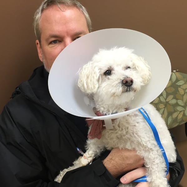
As demonstrated in Daisy's story sprinkled throughout this article, her family got her to the vet quickly, and she recovered well. She was thirteen years old when it happened and she only had one incident.
Please don't take this matter lightly if you suspect your pet has swallowed something and it's gone down the "wrong pipe."