Calcinosis cutis is a rare condition in dogs, and it’s one that can be hard to identify and can take some time to treat.
If your veterinarian has recently diagnosed your dog with calcinosis cutis, we can help you understand what this means and how your vet will help your dog.
This condition needs to be treated by a veterinarian. There aren’t any over-the-counter skin remedies, but there are ways to make your dog more comfortable at home, which we’ll help you with.
What Causes Calcinosis Cutis
Calcinosis cutis is a condition in which the body deposits calcium salts into the skin as hard bumps. This can occur in a single localized area, called calcinosis circumscripta, or more generally across the body, called calcinosis universalis.
Calcinosis Circumscripta
Calcinosis circumscripta often occurs over sites of previous skin damage. It can also occur over spots that commonly experience skin trauma, such as pressure points and bony areas like the elbows, knees, or ankles.
It is especially common in younger dogs because they have extra stores of calcium and phosphorus in their bodies as their bones grow, making it easier to put down calcium salts in their skin when it is damaged. Usually, only one small spot is affected, but some dogs can have multiple calcinosis circumscripta lesions.
Calcinosis Universalis
Calcinosis universalis is the more common form of calcinosis cutis. It is most commonly tied to having extra cortisol in the body. Cortisol is a type of steroid hormone, also called a glucocorticoid. This can occur because a dog is taking glucocorticoids (like prednisone) to treat skin or autoimmune conditions, or when a dog is making extra cortisol themselves due to a condition called Cushing’s disease.
Much less commonly, dogs can develop calcinosis cutis because they have extra circulating calcium in their bodies due to kidney disease, cancer, or because they have eaten a toxin containing high levels of Vitamin D.
If your dog has calcinosis cutis because of extra steroid hormones, they will often have other signs of extra steroids in the body. This can include increased thirst, hunger, peeing, and panting. Additionally, dogs can develop a potbelly, hair loss, muscle weakness, and thinned skin.
What Calcinosis Cutis Looks Like
Pets with calcinosis cutis develop small, hard bumps or flat, raised areas (called plaques) under the skin. These spots are frequently red with gritty yellow crusts on top, and they are often very itchy.
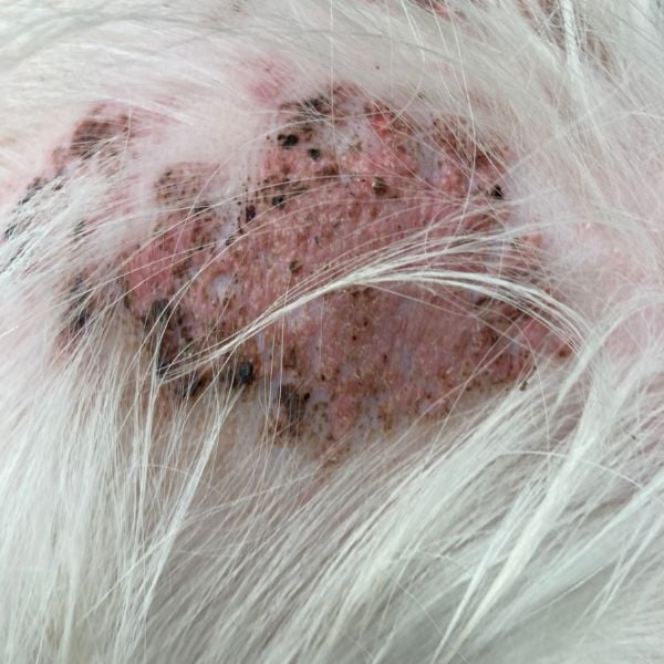
The photo below shows local calcinosis cutis plaque that started a couple of weeks before this dog's appointment. The spots are very red and raised, with small dark brown crusting caused by this dog scratching her neck because the plaques were itchy. Her owner was worried about a new rash on her back after she'd been taking steroids for her allergies for 2–3 years.
In calcinosis universalis, these spots commonly start on the back of the neck or shoulders, but can spread to the entire back and also involve the sides, belly, and face.
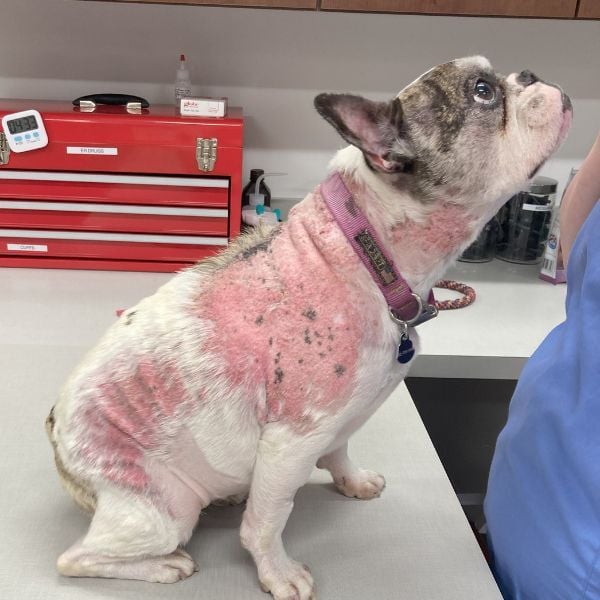
As you can see in the whole body photo of a (cute) Frenchie with calcinosis cutis, there are areas of hair loss on the back and sides, including spreading up to the neck. The bald areas contain raised, bright red calcinosis cutis plaques but not very much crusting.
In the early stages, calcinosis cutis can look like a skin rash on the surface, but it feels very different underneath. If you gently squeeze your dog’s skin around the plaques, they will often feel like jagged plates of armor – inflexible, almost sharp on the edges, and very different from the normal soft skin around them.
A lot of dogs with calcinosis cutis also have skin infections beneath the bumps and plaques, which can look like dark red/brown crusts, skin swelling and tenderness, and small holes on the skin surface that bleed or produce cloudy pink liquid.
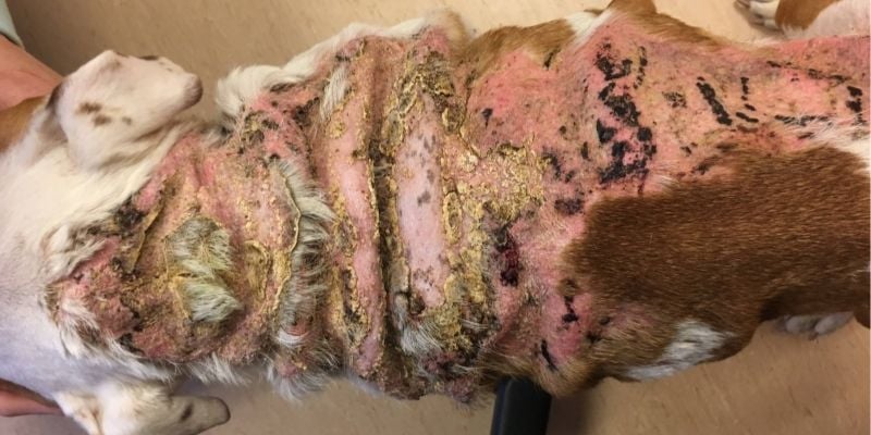
This photo shows a severe case of calcinosis cutis in an English bulldog. The hair loss and red plaques stretch from the back of the neck to the rump, and the yellow-brown thick crusting is caused by infection and extra calcium on the skin surface, along with darker brown crusting caused by his scratching and forming scabs.
Diagnosis for Calcinosis Cutis
Your veterinarian may suspect that your dog has calcinosis cutis based on a physical exam, but they will need to take biopsy samples to be sure.
To take a biopsy, your veterinarian will take several small (5–6mm) punch samples of the plaques and submit them to a pathologist, a doctor who specializes in examining tissue samples. The pathologist will look for evidence of calcium deposits in the skin to confirm your veterinarian’s suspected diagnosis.
Additionally, your veterinarian will want to take surface samples (called a cytology) to look under a microscope for evidence of bacterial infection. If they find bacteria, they may want to send a sample to the lab for a tissue culture to determine which type of antibiotic would work best to treat this infection.
This sample can be collected through a small swab, or they may take an additional punch biopsy for culture. Culture testing can be expensive, but it is very important. Bacterial infections in calcinosis cutis are often very deep in the skin and require sometimes 1–2 months of antibiotics to treat completely. You want to make sure you have the right antibiotic from the start!
If your veterinarian suspects that your dog has Cushing’s disease, they will want to run additional tests to confirm this, including special blood tests (called an ACTH stimulation test or a low-dose dexamethasone suppression test) and an abdominal ultrasound.
Treatment for Calcinosis Cutis in Dogs
If your dog is diagnosed with a single calcinosis circumscripta plaque, which is usually only 1 or 2 inches across, surgery to remove it will cure the condition, and it usually won’t come back. Larger plaques likely won't be removable, especially in areas like the legs if there isn't a lot of extra skin.
If your dog is diagnosed with more widespread calcinosis cutis, the most important part of treatment is identifying and treating the underlying cause. Until this occurs, your dog’s body will continue to put down extra calcium in their skin, and their plaques will get worse.
A dog with Cushing’s disease will need to start treatment to control their extra cortisol production, and a dog taking steroids will need to stop their medications and change to a non-steroid drug to control their other disease.
Often, your dog will continue to form new calcium plaques for at least a few weeks after the underlying cause is addressed, because it takes time for extra steroids to leave their body.
Medications
In addition to the treatments mentioned above, there are also a few medications that can help calcium plaques dissolve faster. The most common treatments are a gel called DMSO (dimethylsulfoxide) and an oral medication called doxycycline or minocycline. These drugs can help your dog’s body start to reabsorb the calcium from their plaques to shrink them.
However, the calcium that’s reabsorbed has to be removed from the body through the kidneys, so it’s vital to have regular blood and urine samples taken by your veterinarian to make sure your dog’s kidneys aren’t hurt by this process.
If your dog has a skin infection under or on top of their calcium plaques, you will need to treat them with antibiotics. These infections can take 1 to 2 months, or sometimes more, to completely heal because they can be quite deep in the skin.
Your veterinarian may also try using a Phovia® light, which uses a special gel and an LED light to emit healing wavelengths into the skin. Phovia can help treat skin infections in dogs with calcinosis cutis, and it can also decrease their skin inflammation to make them more comfortable.
Unfortunately, if your dog’s calcinosis cutis has been present for a long time before they are treated, their spots may never completely go away. The remaining scarred areas usually look a little strange, but your dog should be comfortable and free of itching or pain.
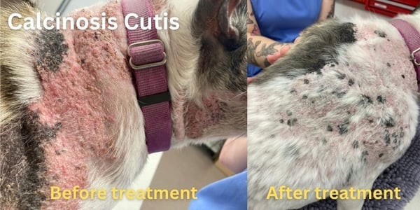
In the photo below, the Frenchie featured in the photos above, is 3 months into treatment — successfully treating with doxycycline and getting the underlying disease controlled. While the plaques haven't completely resolved, they are no longer red, irritated, or crusted, and they show a lot of hair regrowth even though there is still patchy baldness. This would be considered successful healing for a dog with chronic calcinosis cutis, and they will likely continue to heal slowly over the coming years as more calcium is absorbed by the body.
The good news is that you can stop your dog’s body from making new spots by treating the cause of the calcinosis cutis, and you can help break down the plaques partially so your dog is comfortable and unlikely to develop new infections. Your dog’s skin may be partly bald around the plaques, but they will often grow much of their hair back, and their redness and itching should go away with treatment.
Regular bathing (often once or twice a week) can help remove surface crusting and soothe your dog’s itching. Your veterinarian will be able to recommend either an oatmeal shampoo or an antimicrobial shampoo, depending upon whether they find an infection on your dog’s skin. Additionally, you can have your dog wear a t-shirt or other covering to stop them from damaging their skin further while they’re still itchy. Your veterinarian may also be able to recommend non-steroid anti-itch treatments to make your pet more comfortable while they’re in the early stages of treatment for calcinosis cutis.
Working with your veterinarian to identify and treat calcinosis cutis and its causes is the best way to make your dog as comfortable and healthy as possible for all the good times to come.